An In-Depth Look at Lumpy Skin Disease (LSD) in Cattle
Take control of your farm like never before! Say goodbye to paperwork and hello to freedom. Download our apps now!
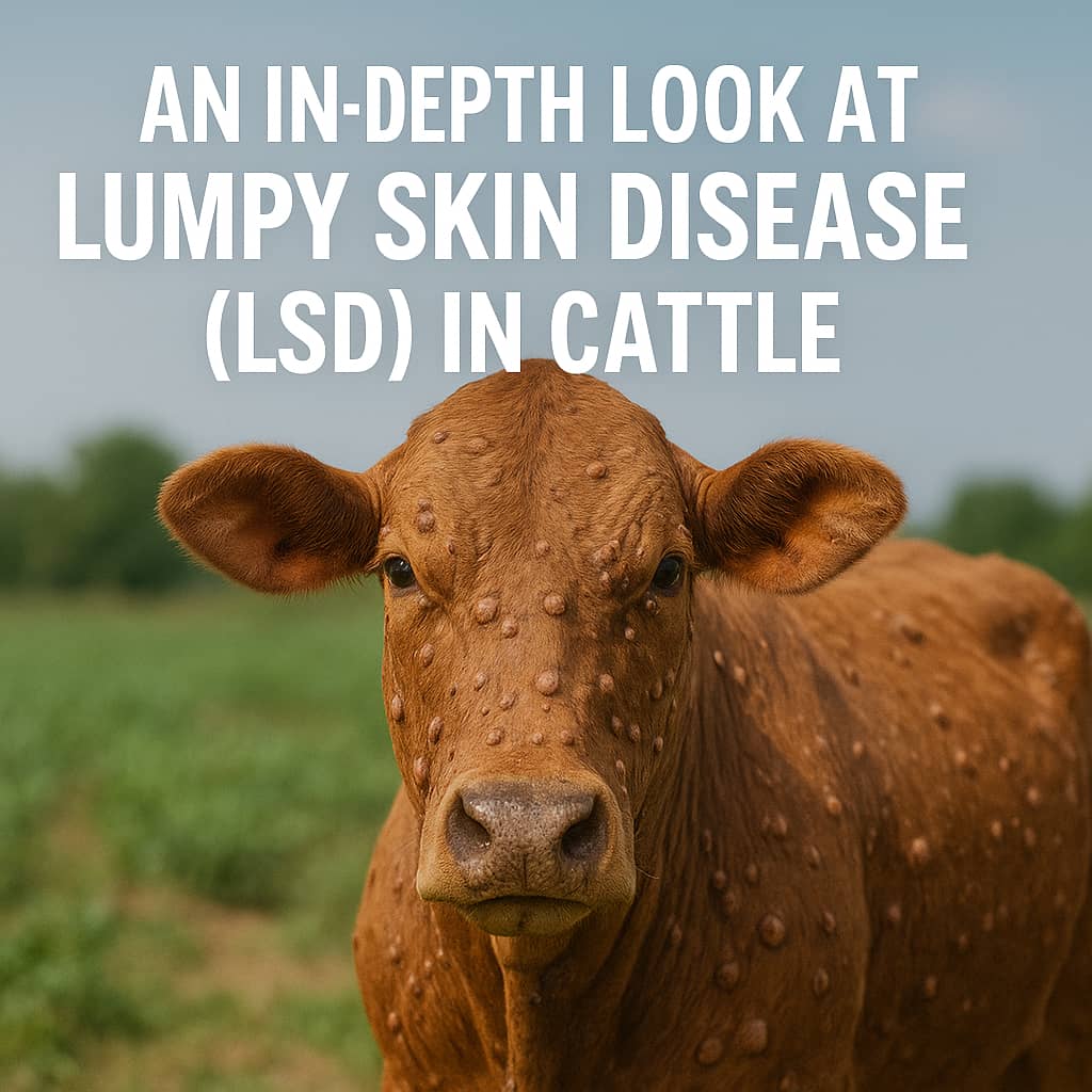
Lumpy skin disease is an acute infectious disease of cattle characterized by the eruption of large numbers of cutaneous nodules and superficial lymphadenitis. The disease has been reported in many parts of Africa.
The incubation period for lumpy skin disease is between 4 and 14 days post-infection. After an initial period of high fever (41°C) and swollen lymph glands, the animal may develop large, firm nodules that are up to 5 cm in diameter in the skin. These can be found all over the body, particularly on the head.
Causes of Lumpy Skin Disease in Cattle
Lumpy skin disease (LSD) is a devastating illness in cattle and buffalo that is caused by a Capri pox virus.
There are two forms of the disease: the Neethling Poxvirus form, which is the true Lumpy skin disease, and the Allerton herpes virus form, which causes a milder version of the disease.
The Neethling virus is responsible for the severe form of the disease, and it is related to the pox virus found in sheep. Initially, the intestinal form of the disease causes constipation, followed by profuse bloody diarrhea, minor ocular lesions, excoriation of the muzzle, and erosions at the commissures of the mouth.
Postmortem examination reveals buccal and nasal lesions, erythema, extensive inflammation and erosion, and muzzle scabs. The carcass is usually emaciated and dehydrated, with skin lesions producing lymph exudation.
The superficial Lumpy skin disease form is characterized by swelling, and the pharynx and larynx sometimes exhibit erosions and ulcerations.
Erythema and edema of the abomasum, as well as internal Lumpy skin disease of the small intestine, are not always present.
Diagnoses of Lumpy Skin Disease in Cattle
To diagnose lumpy skin disease, samples of affected tissues and organs are taken for laboratory examination in 10% formalin (Histopathology) based on the presence of nasal, ocular, and oral lesions, persistent high temperature, and enlargement of superficial lymph nodes.
Additionally, the detection of viruses in lesions can also aid in the diagnosis of lumpy skin disease. Diagnosis can be further supported by the detection of antibodies in serum.
To collect samples for diagnosis, blood should be collected from live animals with a fever. Virus isolation, polymerase chain reaction, and dot blot hybridization can be used to diagnose the disease.
In clinically infected, fevered, and normal dairy cows, lumpy skin disease can also be diagnosed using indirect enzyme-linked immune or bent assay.
The Necropsy Findings of Lumpy Skin in Cattle
In tropical regions, we can use postmortem findings to diagnose lumpy skin disease in cattle. These findings include extensive grayish-pink skin nodes with caseous necrotic centers.
Similar nodes may be seen in the nasopharynx, trachea, bronchi, lungs, abomasums, rumen, renal cortex, testicles, and womb, as well as hemorrhages. Swollen and congested lymph nodes with petechial hemorrhages may also be present.
The differential diagnoses for lumpy skin disease in cattle include Rinderpest, Mucosal disease, and Foot and Mouth disease.
Note that all of these diseases have stomatitis lesions but not on the foot or udder. Prior inoculation with the virulent sheeppox virus can protect cattle against lumpy skin disease.
The virus is present in the blood and saliva and can be easily transmitted from skin lesions. Even when salted, the hide with skin lesions can harbor the virus for up to one month.
Transmission of Lumpy Skin Disease in Cattle
The transmission of Lumpy skin disease in cattle is not entirely clear, but evidence suggests that arthropods, particularly mosquitoes, are the primary vectors. The virus is mainly spread through the bites of flies and mosquitoes, and possibly through ticks.
There is no evidence of transmission through feeding and drinking troughs by saliva. Furthermore, no transmission, whether direct or indirect, has been observed between infected and susceptible animals in experimental settings.
Lumpy skin disease is a viral illness that affects cattle and is primarily transmitted through blood-feeding insects such as certain species of flies and mosquitoes, or ticks. This disease can cause fever and nodules on the skin and can be fatal, particularly in animals that have not previously been exposed to the virus.
Clinical Signs of Lumpy Skin Disease in Cattle
Lumpy skin disease has an incubation period of 5–9 days, after which it is characterized by mild fever and the sudden appearance of skin nodules on the face, neck, back, and perineum.
The morbidity rate is between 5-45%, and mortality typically occurs within 4-14 days, with a low rate of about 5%.
Secondary bacterial infections are possible and can have a high motility rate.
Other symptoms include fluctuating fever, anorexia, increased lachrymation, clear nasal discharge, and swelling of the lymph nodes.
The nodules are round and raised areas measuring between 1cm-5cm, with mouth lesions also possible. The skin nodules may indurate and persist for years, or they may rupture, coalesce, and form wounds.
In some cases, there may be edema of the dewlap and udder. Limbs may also swell, causing lameness.
In cases of Allerton virus infection, mild fever and skin nodules are also present, along with conjunctivitis and excessive salivation.
A drop in milk production, abortion, reluctance to move or eat, and swelling of the limbs, brisket, and genitals may also occur.
Postmortem findings of lumpy skin disease in cattle: Neethling lesions involve all the skin layers and subcutaneous tissue, generalized lymphadenitis, and occasionally erosions & ulcers of the wall of the rumen and abomasums may be seen.
Diagnosis is by histopathology, virus isolation, and clinical signs of the skin lesions, and difficult to differentiate Allerton from the Neethling virus.
Differential Diagnosis of Lumpy Skin Disease in Cattle
When making a differential diagnosis of lumpy skin disease in cattle, skin conditions such as demodicosis and onchocerciasis should be considered.
Treatment of Lumpy Skin Disease in Cattle
Regrettably, there is no direct antiviral treatment available for lumpy cow skin disease. Infected animals are provided supportive care that includes antibiotics, painkillers, and wound care sprays to manage their symptoms. Since no cure exists, vaccines are utilized to prevent disease transmission.
Prevention of Lumpy Skin Disease in Cattle
It is difficult to stop cattle from being attacked by infected vectors like flies and in that case, once the infection is within the area, risk behaviors increase the probability of infection being carried between different locations.
Control of Lumpy Skin Disease in Cattle
The control of lumpy skin disease in cattle is based on four tactics: movement control (quarantine), vaccination, slaughter campaigns, and management strategies.
Annual vaccination with Neethling virus attenuated in lamb kidney tissue or Sheeppox virus tissue culture vaccination can be effective in producing immunity and preventing the spread of disease. However, the use of Sheeppox virus vaccination is limited to countries where sheeppox already occurs.
It is important to note that specific national control plans may vary between countries, and advice from relevant authorities and veterinarians should be sought.
Many people or farmers may mistake lumpy skin disease for other conditions, such as mange caused by the Psoroptes species mites. Mange can cause severe skin disease, including scab formation along the back, shoulders, and tail heads of cattle, as well as intense itching of the skin, weight loss, and death.
Inflammatory responses may result in the formation of crusts and scabs on the skin.
Other conditions that may be mistaken for lumpy skin disease include demodectic mange, insect bites, trauma, urticaria, and photosensitization.
Photosensitization may occur as a primary condition following ingestion of plants like ranunculus bulbosus (buttercup), which contain photosensitizing agents, or as a secondary condition resulting from liver damage that causes the retention of photosensitizing agent phylloerythrin, which affects the skin.
However, these conditions can be differentiated from lumpy skin disease with the help of veterinarians.
Conclusion
In conclusion, lumpy skin disease is a highly infectious and devastating illness that affects cattle. It is characterized by the eruption of large nodules on the skin and swollen lymph nodes. The disease is caused by the Capri pox virus, specifically the Neethling Poxvirus form. Diagnosis of lumpy skin disease involves laboratory examination of affected tissues and organs, as well as the detection of antibodies in serum.
Prevention and control measures for lumpy skin disease include movement control, vaccination, slaughter campaigns, and management strategies. Vaccination with attenuated virus vaccines can provide immunity and help prevent the spread of the disease. It is important to consult with relevant authorities and veterinarians to develop specific control plans for each region.
It is crucial to differentiate lumpy skin disease from other similar conditions, such as mange and photosensitization, as they require different treatment approaches. Seeking professional veterinary advice is essential in accurately diagnosing and managing lumpy skin disease in cattle.
Remember, early detection, proper management, and adherence to preventive measures are key in protecting your cattle from the devastating effects of lumpy skin disease.
Join Our Community ()
Imagine a farm where everything runs smoothly. Make it real—download our revolutionary apps now!
