Mastitis in Cattle: Causes, Signs, Treatment & Prevention
Transform your farm into a well-oiled machine! Streamline tasks, save time, and focus on growth. Download now!
Bovine mastitis is an inflammatory response of the udder tissue in the mammary gland, which can be caused by physical trauma or a microorganism infection. It is prevalent in almost all mammals, but it holds major economic importance in the cattle/dairy industry because it reduces milk yield and affects the quality of milk. Other mammals such as sheep, goats, horses, and camels can also be affected.
The condition is characterized by pathological changes in the mammary gland that significantly impact the quality and quantity of milk produced.
Mastitis can be divided into lactational and non-lactational mastitis, which refer to inflammation of the breast tissue during and outside of lactation.
In a dairy herd, mastitis may follow the usual pattern of infectious diseases, particularly when it is caused by the specific microorganism Streptococcus agalactiae.
Alternatively, it may occur sporadically in one or a few animals in a herd due to a variety of identified microorganisms. Additionally, it may not be associated with any microorganism.
Causes of Mastitis Disease in Cattle
Mastitis is a common udder infection in dairy cows caused by bacterial contamination during milking or environmental contact. Examples of contamination sources include milking equipment, milking personnel, manure, or dirty stalls.
Many microorganisms have been linked to mastitis, which can be classified into two groups: infective and potentially infective. Infective microorganisms, such as Streptococcus agalactiae, are normal inhabitants of either the udder or milk (Staphylococcus aureus) or of the animal (Streptococcus and Staphylococcus) and are responsible for about 1/4 of all cattle cases.
Potentially infective microorganisms, such as Streptococcus dysgalactiae, Streptococcus uberis, and other streptococci, gram-negative rod-like microorganisms, Corynebacteria, pleuropneumonia-like types, and those having characteristics of yeast, are found in the animal's environment, such as dung, urine, and bedding, and can cause mastitis when the animal's defense mechanisms weaken.
Transmission of Mastitis Disease in Cattle
It primarily spreads during milking via the milking machine, contaminated operators' hands, and materials (such as cloths) used to wash the udder. Mastitis occurs when microorganisms enter the udder through the teat canal.
There are various predisposing factors for mastitis in cattle. The older the animal, the greater the likelihood of infection. Mastitis is also more likely to occur at the start and end of lactation, and high milk yielders are more often affected. Hereditary factors, such as low-slung udders and long teats, can also make mechanical damage more likely and allow for invasion more quickly.
Hygiene measures can be taken to reduce the number of microorganisms and decrease the chances of infection. However, there may be unknown factors that make it possible for two identical animals in the same environment to contract the disease, while one is completely resistant. The method of milking, such as stripping, can also increase the chances of infection.
There are three forms of clinical signs of mastitis in cattle: subclinical mastitis, mild clinical mastitis, and acute/severe clinical mastitis.
a). Subclinical Mastitis
Subclinical mastitis can only be identified by laboratory examination of milk drawn from the udder. There are no visible signs, and the milk drawn is also normal. Many cases of subclinical mastitis are due to Streptococcus agalactiae.
b). Mild Clinical Mastitis
Mild clinical mastitis is characterized by distinct changes in the udder, which may be detectable by palpation. The organ is firm to the touch in one or more teats, and there are changes in milk consistency, such as watery or abnormal color (often pink or yellow due to blood stains) and flakes or clots (large or small), indicating inflammation. Milk from one teat may increase, while one or more teats may be affected, while the others remain normal.
c). Acute/Severe Clinical Mastitis
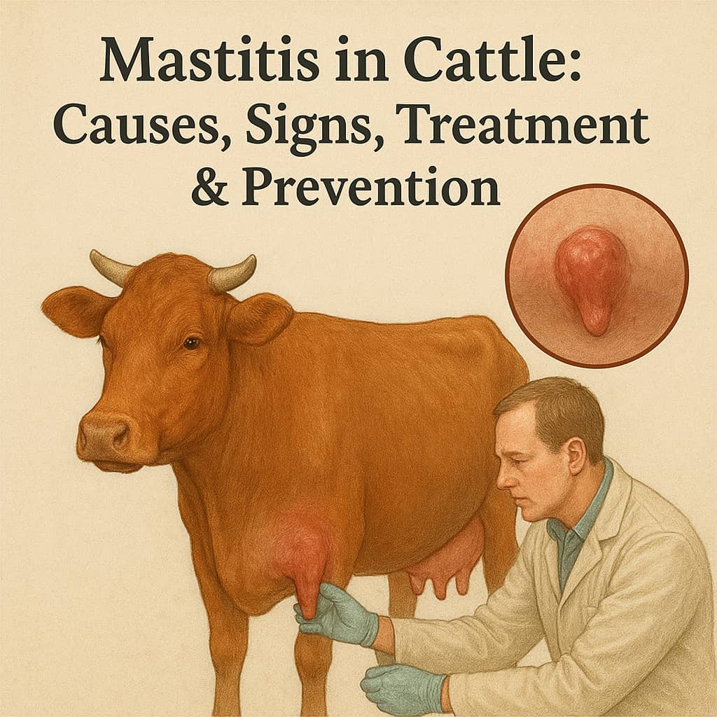
Acute/severe clinical mastitis is characterized by obvious symptoms of clinical mastitis in the udder, such as swelling, heat, hardness, redness, or pain. Milk takes on a watery appearance, and flakes, clots, or pus are often present.
Milk changes may also include a precipitate of yellow sediment, clear yellow streams with or without blood clots and white/yellow flakes, pink color with or without sediment, or green or yellowish-green sediment with a foul smell (when the infection is due to Corynebacterium pyogenes).
The quantity of milk drawn is usually small. The animal may show a degree of systemic reaction, such as increased temperature and pressure, anorexia, and arching of the back, and may have difficulty rising. Teat examination may reveal injuries or sores, especially near the orifice.
In chronic mastitis, which follows acute mastitis, there is almost no pain. The udder progressively indurates and then atrophies. The animal may eat normally and not have a fever, but milk secretion gradually decreases.
Diagnosis of Mastitis in Cattle
The use of the strip cup is highly recommended as it provides the initial indication of mastitis presence. A strip cup is a metal container with a cup-shaped design and a 250ml capacity. It has a ledge of approximately 3cm down from the rim, where a fine gauze disc is placed.
The milk stream is directed onto the gauze, which holds any flakes and clots. Another option is a shiny black top plate that can be used instead of gauze. This alternative can identify discoloration as well as other abnormalities. After using the strip cup, suitable treatment can be administered.
Although laboratory testing can be conducted, contamination during the collection of samples is a common occurrence that can significantly alter the results. To mitigate this risk, it is important to follow the procedures outlined below;
- Wash the teats and dry them (no water in the collecting tube).
- Clean the teat orifice with cotton wool dipped in spirit, squeezing the end of the teat while cleaning to extrude the wax plug and dirt normally found around the orifice.
- Direct the first few squirts/streams into the sterile bottle.
- Label the bottle with the animal number and quarter.
- Keep samples cool (put them in ice).
- Forward to the laboratory for examination.
- Use the California mastitis (Schalm test).
- The test makes use of the fact that mastitis milk contains more cells, mostly leucocytes, than normal. If this is mixed with agents like alkyl-sulphonate, a gel is formed owing to a reaction between the test solution and the DNA of leucocytes. The 4 quarters are milked (discard the first stream).
Simple Testing of the PH of Milk
- Use mastitis detector blotters. These are sheets of thick bloating paper, about 10cm
with 4 circular areas, each 2cm in diameter. These contain bromothymol blue and are yellow.
- Direct the 2nd stream from each quarter to each area in turn. Milk from the infected quarter turns yellow with a light or dark - greenish-blue color depending on alkalinity.
- Normal milk has a PH of 6.6. With mastitis, it tends to increase towards that of blood-7.4.
- This test is not very reliable in cows at the beginning and end of lactation because milk in these animals is more alkaline.
- The test is done on a plate consisting of shallow cups. An equal amount of test solution is added to each. Mix by rotating the paddle in a circular motion.
- Normal milk remains liquid. The gel forms (for mastitis milk) in the center of the cup.
Note: The pH of milk accounts for the amount of lactic acid produced by microbial activity. The more lactic acid present, the higher the acidity. This would result in a change in taste and smell, making it unsuitable for human consumption.
Treatment of Mastitis in Cattle
Once mastitis has been detected, first aid involves applying ice cubes to the surface of the udder. The infected milk from the affected teat should be drained three times a day and safely disposed of. A solution containing 5% phenol can be added to the infected milk to ensure proper and hygienic disposal.
Antibiotics such as cephapirin, pirlimycin, and ceftiofur are administered through the udder infusion via the teat canal. Before inserting the nozzle of an intramammary tube, the teat orifice should be cleaned with cotton wool soaked in spirit to prevent the entry of extraneous microorganisms.
Antibiotics Used in The Treatment of Mastitis
The use of penicillin can lead to the development of resistance over time, necessitating a switch to another drug. Additionally, residual penicillin in milk can interfere with the manufacturing process and pose a health risk to those who consume it. It should be noted that not all cases of mastitis respond to penicillin, but Streptococcus agalactiae and Staphylococci pyogenes are among the microorganisms that do.
For coliform infections, Streptomycin and Tetracycline are recommended, while Neomycin and Polymixin are used for Pseudomonas infections. Erythromycin, a broad-spectrum antibiotic, is often used when the causal organism is unknown.
Recommended dose rates by intra-mammary infusion.
- Penicillin: apply 50,000 to 200,000 units.
- Tetracycline: apply 100 to 400mg.
- Streptomycin: use 0.25 to 1.0g.
- Polymixin (B.Sulphate): use 50mg.
- Erythromycin: use 300 to 600mg.
Note: In acute systemic mastitis, systemic injection of antibiotics is necessary.
How to Control Mastitis in Cows
Mastitis can be treated with intramammary or systemic antibiotics or a combination of both. Intramammary drugs are generally most effective for mild mastitis in a single quarter, while systemic treatment is preferable for severe cases or infections affecting multiple quarters.
To prevent the spread of mastitis, it is important to implement frequent testing, thorough treatment, culling of infected animals (especially those that are not the best in the herd), and proper hygiene practices. This includes:
- Milkers should wash their hands thoroughly before and after milking. Those who are sick, especially with a sore throat, should not milk.
- Clean clothing should be used, and a separate clean towel should be used for each cow. The towel should be rinsed in disinfectant before and after use.
- The first stream of milk should be discarded and not allowed to touch the floor.
- Milkers should not wet their hands with milk or saliva.
- Increase the amount of water used for washing after milking, and teat dipping should be done within a few minutes of milking.
Teat disinfection serves three functions, including killing bacteria that may be transferred by the milker's hand or machine to prevent the establishment of a bacterial colony at the teat end.
When mixed with an emollient, it keeps the teat supple and prevents chapping and other lesions that could harbor S. aliens and Streptococcus dysgalactiae.
Teat chaps with bacteria growing in them take a long time to heal; therefore, the antiseptic properties of teat disinfectants also promote the healing of teat lesions.
Dry-Cow Therapy (DCT)
A dry cow is a cow that hasn't been milked for a couple of months. This is an intentional rest period given to all dairy cows to allow them to recover and recuperate. The treatment and management of dairy cows during this period of inactivity is called Dry Cow Therapy (DCT).
DCT involves the infusion of a specially formulated long-lasting antibiotic into each quarter of the udder. This serves to eliminate any remaining infections and prevent new ones during the dry period. Preparations containing Cloxacillin are very effective.
The dry period enables the cow and her udder to recover and regenerate. The goal of dry-cow therapy is to increase the chances of a cow giving birth with a low somatic cell count (SCC), meaning that the cow is not infected, and reduce the risk of developing clinical mastitis during the next lactation.
Conclusion
In conclusion, bovine mastitis is a significant issue in the cattle/dairy industry due to its impact on milk yield and quality. It is an inflammatory response of the udder tissue in the mammary gland, primarily caused by microorganism infections or physical trauma.
The condition can be categorized into lactational and non-lactational mastitis, occurring during and outside of lactation, respectively. Various microorganisms, both infective and potentially infective, have been linked to mastitis, with bacterial contamination during milking or environmental contact being a common source.
Mastitis spreads through milking, where microorganisms enter the udder through the teat canal. Predisposing factors such as age, lactation stage, high milk yield, and hereditary factors can increase the likelihood of infection. Hygiene measures during milking and reducing the number of microorganisms in the environment can help prevent mastitis, although there may be unknown factors influencing its occurrence.
Clinical signs of mastitis range from subclinical (only detectable through laboratory examination) to mild clinical (changes in udder firmness and milk consistency) to acute/severe clinical (obvious udder symptoms, changes in milk appearance, and systemic reactions). Diagnosis involves the use of strip cups, laboratory testing, and pH testing of milk.
Treatment includes applying ice to the udder surface, draining infected milk, and administering antibiotics via udder infusion. Different antibiotics are used based on the causative microorganism, and proper hygiene practices are crucial to prevent the spread of mastitis. Control measures involve testing, thorough treatment, culling of infected animals, and hygiene practices during milking, including handwashing, clean clothing, and teat disinfection. Dry-cow therapy during the resting period helps eliminate existing infections and reduce the risk of clinical mastitis in the next lactation.
Effective management strategies, early detection, and appropriate treatment protocols are essential for controlling and minimizing the impact of mastitis in cattle, ensuring the production of high-quality milk in the dairy industry.
Join Our Community ()
Imagine a farm where everything runs smoothly. Make it real—download our revolutionary apps now!
